
Joint Surface Damage
The bony surfaces of the knee joint are lined by spongy (hyaline) cartilage. This provides cushioning to the underlying bone and provides a near frictionless surface for the joint to move. This hyaline cartilage can be damaged causing an isolated cartilage defect or if the underlying bone is affected an osteochondral defect.
Damage to the cartilage can be the result of a traumatic injury or from generalised wear and tear without any history of trauma.
Symptoms
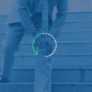
The majority of patients complain of a dull aching pain that is made worse with exercise and relieved with rest. This can also be associated with intermittent knee swelling, joint instability and catching pain on certain knee movements.

Treatments
Non-operative
Not all cartilage defects require surgery. Defects can heal without surgery especially in younger patients. Use of anti-inflammatory medication, intraarticular steroid or viscosupplementation injections and physiotherapy can help improve symptoms.
Operative
If non-operative treatment fails or if the defect is causing significant symptoms to the knee, arthroscopic (keyhole) surgery is recommended. This type of surgery includes:
- Debridement/chondroplasty – This is performed if the cartilage defect is of partial thickness. The unstable flaps of cartilage are shaved back during the arthroscopy to stable edges and thereby smooth the joint surface and remove loose fragments. This is a day case procedure and after surgery there are no mobility restrictions.
- Microfracture – This is performed if the cartilage defect is full thickness, it is a procedure to try to restore host cartilage. Small drill holes are made within the bone to allow blood and stem cells to coat the area of the defect and initiate a healing response. This is a day case procedure. After surgery a hinged knee brace will be supplied to allow a full range of motion. For 6 weeks after the surgery you will be mobilising non weight bearing with crutches. It takes up to 12 weeks for the cartilage to repair, therefore the rehabilitation back to full sports is generally 4-5 months after surgery.
- Internal Fixation – This is used for large osteochondral fragments where the fragment is either still attached to its original position or is loose. Fixation of the fragment back to its original site can be performed to enable it to heal. This is a day case procedure. After surgery a hinged knee brace will be supplied to allow a full range of motion. For 6 weeks after the surgery you will be mobilising non weight bearing with crutches. It takes up to 12 weeks for the fragment to heal, therefore the rehabilitation back to full sports is generally 4-5 months after surgery.
- Cartilage Regeneration – For larger cartilage defects where microfracture on its own is unlikely to work a collagen matrix graft is glued into place in the defect. Through the microfracture drill holes stem cells leach out and are contained within the graft where they can initiate a healing response. This is a day case procedure. After surgery a hinged knee brace will be supplied to allow a full range of motion. For 6 weeks after the surgery you will be mobilising non weight bearing with crutches. It takes up to 12 weeks for the cartilage to repair, therefore the rehabilitation back to full sports is generally 4-5 months after surgery.